The content of the article
Fibroadenomas are often detected by chance with an independent examination of the mammary glands or with ultrasound, mammography. These are small dense nodules that are located in the thickness of the breast tissue. In the classification according to ICD-10, the disease passes under the code D24.
Reasons for the development of pathology
So far, the exact causes of the emergence and development of such formations have not been identified. But it is known that this is the result of hormonal disorders. Estrogens are responsible for the growth of breast tissue, the formation of new lobules. Progesterone is a kind of estrogen blocker, it stimulates the differentiation of formed tissues. Normally, these processes are in equilibrium. When estrogen levels are elevated and progesterones are lowered, foci of hyperplasia are formed, which subsequently form fibroadenomas.
An imbalance of hormones occurs against the background of problems with the thyroid gland, malfunctions of the pituitary gland, ovaries, adrenal glands. Such violations are the result of pathologies of the kidneys and liver, malfunctions in the menstrual cycle, diabetes mellitus, obesity, and the influence of adverse external factors.
With the onset of pregnancy, a cardinal hormonal reorganization begins in the body, which contributes to an increase in neoplasms in size and quantity. This process may interfere with normal lactation.
There are periods of risk:
- adolescence;
- menopause;
- the use of contraceptives;
- recovery from a miscarriage or abortion.
Classification
The following types of pathology are distinguished.
- Phyloid (leaf). It is a limited seal, which consists of individual nodes woven together. Often accompanied by painful sensations, which is uncharacteristic of other forms. It grows slowly, so it can go unnoticed for a long time. Enhanced growth is accompanied by abundant discharge from the nipples. With a large tumor, the skin on the chest acquires a purplish-cyanotic shade, stretches, becomes thin, through which the vascular and venous networks are visible. Such a tumor is capable of acquiring a malignant course, but is quite rare.
- Intracanalicular. Connective tissue penetrates into the lumen of the ducts and fits snugly against the walls. The tumor does not have clear boundaries, is characterized by lobular structure and heterogeneous structure. It is not amenable to medical treatment. It’s hard to identify.
- Pericardial. Sites of fibrous tissue grow around the milk ducts. Education is limited from other tissues, has a clear contour and dense structure. Calcium salts can settle in it (especially in elderly women), calcified fibroadenoma occurs.
- Combined. The signs of intracanalicular and pericanalicular are combined. Such a neoplasm grows around the duct and inside it, has a lobed structure and a heterogeneous structure. It does not differ in obvious symptoms, it is very rarely manifested by soreness.With growth, it changes the shape and appearance of the surface of the chest. More often localized in the upper quadrants.
According to the degree of maturity, the following types of fibroadenoma are distinguished.
- Mature. The neoplasm has a clearly formed capsule of dense consistency. It grows slowly, practically does not increase. It is typical for women 20-45 years old.
- Immature. The neoplasm has a soft elastic consistency, prone to rapid growth. Most often found in girls during puberty. It can dissolve on its own as the regulation of the hormonal background or the establishment of the menstrual cycle.
Symptoms
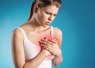
Can an adenoma hurt? Pathology has no characteristic symptoms. You can recognize it by its shape and density during an independent examination and palpation of the chest. Most often, the formation is localized in the upper part. It can appear on both the right and left breasts.
A single neoplasm is represented by an elastic ball with clear borders with a diameter of 1-7 cm. Such fibroadenomas do not change the appearance of the chest. The seal may increase to 20 cm, in which case it will be noticeable as a bulge on the surface of the mammary gland.
Since the pathology is associated with hormonal disorders, it is often accompanied by:
- acyclicity of menstruation;
- sudden loss or weight gain;
- visual impairment;
- fatigue.
In phyloid fibroadenoma, the symptoms are more pronounced:
- chest pain - intensify during menstruation;
- enlargement, change in the shape of the glands - one or both.
Self-examination
Given that the first sign is most often a modification of the breast tissue or the presence of compaction, pathology can be detected by self-examination. Characteristics of neoplasm during palpation:
- resembles an elastic ball;
- has clear contours;
- movable;
- located more often in the upper part of the mammary gland;
- with pressure it does not cause pain;
- with large sizes, changes the appearance of the breast.
If fibroadenoma has just begun to develop and has small dimensions, it is difficult to detect it yourself. Especially when the chest is medium and large.

Diagnostic methods in young women and in menopause
The likelihood of neoplasm formation depends on fluctuations in the hormonal background. For this reason, pathology is often found in young women and those who have reached the threshold of menopause. The earlier fibroadenoma is detected, the easier it is to get rid of it. It is important to visit a mammologist regularly. For any changes in the mammary gland that are visible visually and to the touch, inform a specialist.
The following diagnostic methods are used to confirm the diagnosis.
- Inspection. Careful palpation of each area of the mammary gland allows to reveal the formation of even small sizes. The smaller the size of the breast, the easier and more informative the examination. It is also easy to detect nodes localized near the nipple.
- Breast ultrasound. It is most informative to perform in women of reproductive age - up to 45-50 years. It allows you to differentiate the neoplasm and gives an idea of those areas and tissues that cannot be seen during x-ray examination. But it does not reflect the nature of the tumor - malignant or benign.
- Mammography. Suitable for menopausal women. In the reproductive period, glandular tissue will not give an informative picture, and in menopause, the mammary glands are represented by adipose tissue, which does not absorb x-rays. Mammography is also informative in tumor calcification, since calcium salts are X-ray positive.
- Biopsy. With fibroadenoma, a puncture biopsy is performed, in the presence of a cyst - aspiration. Make a fence of a certain amount of tissue for the purpose of further histological study.This allows you to establish the nature of the tumor, the degree of tissue damage. It is performed on an outpatient basis under local anesthesia. In fact, this is an “injection” into the tissue of the mammary gland. Ultrasound is used for accuracy. If the results of the fence are doubtful, an additional trepan biopsy is performed using special thin needles that have a thread. They are "screwed" into the tissue, and then sharply removed. As a result, larger particles remain on the thread than with a conventional biopsy.
- MRI and CT. They help to differentiate with cysts, malignant tumors, to study regional lymph nodes.
Types of conservative treatment
Treatment without surgery is permissible in the following cases:
- the size of the formation does not exceed 2 cm;
- there is no upward trend;
- no complaints from a woman;
- a tumor detected during puberty;
- the type of fibroadenoma is not leafy.
A woman should be monitored regularly by a mammologist. It is recommended to normalize body weight and adhere to a healthy diet.
Hormone replacement therapy is mainly prescribed. Homeopathic remedies, preparations with a high content are also used. vitamin e and iodine, immunostimulants for the complex effect on the problem.
Operations
Radical removal is possible only surgically. This does not affect subsequent breastfeeding. However, even after such treatment, the woman is not safe from tumor recurrence, but in a different place. Indications for the operation are as follows:
- the size of the tumor is more than 2 cm;
- phyloid type (absolute indication);
- pregnancy planning, including IVF.
During gestation, surgery is not performed.
The tumor is removed in several ways.
- Sectoral resection. The tumor is removed along with a piece of glandular tissue of the lobule where it is located. Such an operation does not lead to asymmetry and deformation of the chest. It is carried out mainly under general anesthesia. The method is used for suspected malignant degeneration, diffuse fibromatosis, large tumor sizes.
- Enucleation. By husking, only fibroadenoma is eliminated without the removal of surrounding tissue. During this kind of surgical intervention (using a scalpel), tissue is taken for subsequent histological examination. The treatment is easily tolerated, the postoperative and rehabilitation periods pass quickly, the seams are almost invisible.
- Laser removal of nodes. A special conductor is brought aiming at the fibroadenoma, then a laser is fed through it. Education "evaporates." Cosmetic defect and consequences are minimal.
- Cryoablation. It provides for the supply of liquid nitrogen to the tumor and "freezing". After this, destruction of the nodes occurs within a few weeks.

Folk remedies
Treatment with folk remedies can only be used in combination with conservative methods or during the rehabilitation period. Below are some recipes.
- Decoction. Used to correct estrogen and androgen levels. Fennel Fruits, Flowers daisies, wheatgrass roots, licorice and Althea unite in equal parts. One tablespoon of this mixture must be filled with a glass of boiling water and infused for 15 minutes. It is taken in strained form three times a day for three weeks. A new broth is prepared daily.
- Infusion. Used to slow the growth of tumors. Three teaspoons of wormwood herb must be poured with a glass of boiling water, placed in a thermos and insisted for three hours. Pass through cheesecloth and drink a teaspoon twice a day.From the third day of admission, increase the dose to one tablespoon twice a day. The course of such therapy is ten days.
- Ointment. It is used to reduce the size of education. 200 ml of refined sunflower oil must be combined with a small piece of yellow wax in a metal bucket and put on fire. After the wax melts, add pre-cooked and chopped chicken egg. Boil until foamy. Then remove from the stove, wait until it subsides, and then again keep it on low heat for 30 minutes. Strain the mixture and let it brew for several hours. Apply to the chest with light movements twice a day.
Signs of cancer
Only a specialist can differentiate a benign neoplasm from a malignant one. But there are signs by which to distinguish fibroadenoma from cancer. The main criteria are presented in the table.
Table - Signs by which fibroadenoma can be distinguished from cancer
| Criterion | Fibroadenoma | Crayfish |
|---|---|---|
| Tumor growth | Slow | Fast |
| Circuit | Smooth | Hilly |
| Consistency | Dense | Dense |
| Attitude to surrounding tissues | Isolated | Soldered with them |
| Mobility | Easy to shift | Limited |
| Pressure pain | Not | Is there or not |
| Axillary lymph nodes | Not increased | May be increased |
Prevention
Measures to prevent the appearance of pathology are aimed at creating conditions that support a normal hormonal background. To do this, you must:
- lead a healthy lifestyle - and also eat right, maintain immunity;
- protect yourself from excessive loads - physical and emotional;
- treat endocrine diseases on time - and periodically inspect your body for changes.
Fibroadenoma is always associated with hormonal imbalance. Most often, its removal is carried out with subsequent histological examination of tissues. Conservative treatment is effective only with small sizes and subject to regular medical supervision. You can see education in the photo on the Internet.
Reviews
I was removed from both sides at the age of 21 (now 28), local anesthesia. They did it in the morning, and already wrote out in the evening))) the scars on the areole go, not at all noticeable. Now I breastfeed the baby))) so everything is ok. The operation is simple, do not be afraid. Good luck and health!
a guest, http://www.woman.ru/health/woman-health/thread/3857900/
In 1991, they removed the fibroadenoma, and besides, there were just two in both breasts, it was poorly tolerated, and local anesthesia painfully departed. I was 18 years old then. After there were childbirth and abortion, here after the abortion I again had fibroadenoma, they offer surgery again, but I refuse, I'm afraid. It doesn’t bother me, it has not been increasing for 12 years now. Only, of course, one should be observed by a mammologist. I wish you all good health!
Natalya, http://www.woman.ru/health/woman-health/thread/3857900/
I had fibroadenoma at age 18 (now 25). Did a puncture, showed that it requires removal. We performed the operation under local anesthesia, the operation went fine, there is nothing terrible here (you don’t feel pain at all). But after the operation, when the anesthesia goes away ... the pain is terrible. But everything passes, is forgotten.
At 21, she gave birth to her first child, there were no breast problems at all. At 24, she gave birth to a second, after childbirth milk stagnation formed in the scar site, but the geneticist said to not touch and everything normalized in a day, it happened (but honestly, I was very worried ... I thought maybe I could do the massage ... but I obeyed the doctor and did it right) . So there is nothing terrible here !! I gave birth to two children, the first fed 1 year, the second feed and now (we are half a year). So good luck, everything will be fine !!!katiyra_kz, https://mamochki.by/forum/1/53252/
0 days ago I was on ultrasound of the mammary glands and I was diagnosed with a cyst in doubt.
Yesterday I went to a consultation with an oncologist. there, too, examined, did an ultrasound, took a puncture. eventually diagnosed with fibroadenoma 1.3 * 0.9 cm in size.
gave a form with the names of the analyzes that need to be submitted before the operation. they want to send to the 33rd hospital in falconers. They said they would have to lie down for 2-3 days.
I have a small child, one year old and not, I don’t even know what to do ...
Is it worth rushing to go to bed under a knife? I don’t knowCaramel, http://club.passion.ru/zhenskoe-zdorove/fibroadenoma-mzh-t91658.html
I have, I go with her for 3 years. in the spring and autumn on the control ultrasound, the main thing that would not grow. the doctor said that while it does not increase it is not necessary to remove. The main thing is that there would be no hormonal jumps. special control in case of pregnancy.
MiSteriya, https://www.u-mama.ru/forum/family/health/446568/index.html
