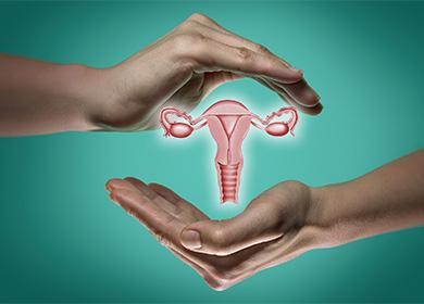
The content of the article
Today, even small-sized tumors are diagnosed without problems - up to 1 cm, which is often a "find" during the examination. Some women can “carry” myoma through their whole lives and not have any problems with it, while others are forced to go to surgery to remove the uterus due to complications.
Tumor features
Myoma is a tumor that is prone to slow growth and has a benign character. Significant fibroids appear not in a year or two. With rapid growth, the malignant nature of the formation cannot be ruled out.
The exact mechanism and causes of the formation of uterine fibroids are unknown. Recently, there has been an increase in the incidence among girls under 30 years old. This is due both to the modern lifestyle and to a high level of diagnosis in gynecology.
The main facts regarding fibroids are as follows.
- Hormone dependent. It happens only in women of reproductive age, and in the menopause it “dries out,” because sex hormones are necessary for its growth. Identification of nodes in women during menopause should always cause a suspicion of a malignant tumor.
- Combined with hormone imbalance.. 80% of women with uterine fibroids have hormonal disorders - irregular menstruation, signs of polycystic, uterine polypsviolation of the thyroid gland.
- "Friend" mastopathy. The mammary glands and the uterus are closely related functionally. Changes in one organ lead to disturbances in another, so most girls with uterine fibroids show mastopathy of varying severity. It is necessary to be treated comprehensively, and not separately, each part of the body.
- There is no cure for fibroids. All the knowledge that doctors have today regarding the mechanism of formation of myomatous nodes does not allow us to identify the true mechanisms of pathology. All existing drugs can only temporarily stop tumor growth and the development of complications.
- It has many synonyms. At different times, this tumor was called differently. The fact is that the uterus consists of myometrium (muscle) and connective tissue. It is impossible to say what tissue the node consists of without removing it and histological examination, relying only on this ultrasound, CT or MRI. This does not affect the tactics of women with such neoplasms.
Histologically distinguish:
- leiomyoma - the tumor consists only of smooth muscle fibers;
- rhabdomyoma - from muscle tissue of another species;
- fibromyoma - muscle + connective tissue;
- fibroma - more than 90% of the tumor consists of connective tissue.
Classification
Myoma is classified as follows.
- Small. If there is one or more nodes with the largest size up to 5 cm.
- Big one. If there is at least one node more than 5 cm or if the uterus, when examined, corresponds to a size of 12 weeks of pregnancy (about 10-12 cm in diameter).
- Multiple (multinodular). If clearly distinguishable three or more nodes.
- Single. If there is only one node.
- Submucous (submucosal). If the tumor is located with a protrusion in the uterine cavity, deforming it.
- Subserous (subperitoneal). If the node "like a mushroom" protrudes above the surface of the uterus into the abdominal cavity.
- Interstitial (internal). If the node is located in the thickness of the muscle wall
- Mixed. Often one node has several growth directions, then they talk about interstitially submucous or subserous-interstitial node.
- Symptomatic The term is used if myoma is the cause of some pathological conditions. Most often it is anemia due to bleeding.
- Cervical. If the node is located in the cervical region (5-7% of the total number of this tumor).
Reasons for education
What is a nascent myomatous node? Specialists can not name an unambiguous reason for the development of pathology. It is believed that against the background of a genetic predisposition, a tissue site with an increased number of sex hormone receptors is formed in the uterus. They are also more sensitive to estrogens and progestogens than similar formations in neighboring cells. As a result, this area begins to grow more intensively compared to the rest. The body loses control over it, a tumor develops, which can reach a diameter of more than 20 cm.
Risk group
Uterine fibroids can occur without obvious causes and predisposing factors. But more often develops in the following women:
- if close relatives suffered from this disease;
- if there is a hormonal imbalance;
- with excessive weight;
- with constant stress, lack of sleep, chronic fatigue;
- nulliparous;
- with metabolic disorders and diabetes;
- after numerous stimulations of ovulation or IVF procedures.
Symptoms
Signs of pathologists are noticeable only when the node reaches a significant size - more than 2-3 cm for submucous and 5 cm for interstitial and subserous. Until this time, a girl can "grow" a tumor for a long time.
- Pain. This is the most common symptom of the disease, which reflects the stage of progression of the disease and the degree of involvement of other organs in the process. The pains are localized in the lower abdomen, in the lower back. May intensify on the eve of menstruation, during sexual intercourse, exercise. As the tumor grows, the pain becomes permanent - it is pulling, there is a feeling of a "stone" in the lower abdomen.
- Bleeding. Abundant periods also often accompany fibroids. They arise for several reasons. For example, if the node is located submucous (submucosal), it interferes with normal endometrial rejection; if the node is large, it deforms the uterine cavity, increases the “bleeding” surface and interferes with the normal contraction of the myometrium; if fibroids are combined with endometrial polyps (this often happens, since both diseases are the result of hormonal disorders). Regular blood loss leads to the development of anemia with their vivid clinical picture - pallor, lethargy, weakness, lethargy, shortness of breath, dizziness.
- Infertility. Small tumors practically do not affect the course of pregnancy. But large or growing in the uterine cavity interfere with the normal development of the fetal egg. Myoma is less extensible compared to normal myometrium, so with the onset of intensive growth of the uterus, the likelihood of miscarriages and premature births increases.
- Constipation. If the fibroid is very large or grows toward the rectum, compression of the latter occurs, which leads to regular constipation.
- Urinary Disorder. A similar pattern occurs with the growth of nodes on the front wall of the uterus, but in this case there is incontinence or frequent urination.
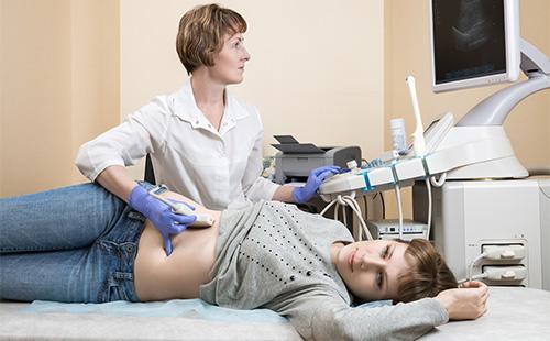
Diagnostics
The most affordable and effective method for the diagnosis and confirmation of fibroids is ultrasound of the pelvic organs. But even with a routine gynecological examination, the doctor may suspect a tumor if an enlarged uterus or its tuberosity is determined.
To identify fibroids, an ultrasound scan must be performed, observing the following recommendations:
- run at the beginning of the cycle - nodes on the third to fifth day have their true sizes, also at this time it is possible to reliably determine the condition of the endometrium;
- use a vaginal probe - it allows you to distinguish even small tumors up to 2 cm;
- track dynamics - to monitor the growth of nodes, it is necessary to conduct an ultrasound regularly every six months.
If submucous nodes are suspected, hysteroscopy is performed, which allows you to immediately remove small-sized formations.
Complications
Often, uterine fibroids are asymptomatic, but in 10-15% of cases, life-threatening complications arise.
Necrosis or torsion of the node legs
The subserous nodes can be displaced about its axis, which leads to compression of their blood vessels and tissue necrosis. In this case, a typical clinical picture of peritonitis occurs. The main symptoms are as follows:
- abdominal pain, often after exercise;
- temperature rise;
- on examination and palpation of the uterus painful;
- in the blood - signs of inflammation;
- ultrasound reveals nodes with signs of degeneration.
Any myomatous nodes are subject to necrosis, even with an intramural arrangement. But if the fibroid has a leg, the probability is higher.
Inflammation
In the presence of chronic inflammation in the pelvic area (e.g., adnexitis, colpitis, endometritis) pathogenic microorganisms can penetrate the blood and lymph vessels to the myoma. The nodule becomes painful due to tissue edema. Symptoms similar to fibroid necrosis appear - pain, temperature, changes in blood tests. The only difference is that against the background of antibiotic therapy, a decrease in all complaints is noted.
"Birth"
Submucous nodes are at least 1/3 protrude into the uterine cavity. If the myoma has such directed growth, over time it “hangs” into the uterine cavity, connecting with myometrium only with the leg. Gradually, the “birth” of such a submucous node can occur - it leaves the uterine cavity first into the cervical canal, and then into the vagina. The woman notes the following:
- cramping pains in the lower abdomen;
- spotting from the vagina.
The treatment in this case is only surgical - removal of the node by twisting or abdominal surgery with a deep leg in myometrium.
Malignancy
In 1-3% of cases, fibroids can degenerate into sarcoma. The main signs of malignant growth are as follows:
- rapid node growth over the past six months to a year;
- the appearance or increase of fibroids in menopause.
The probability of an oncological process in large tumors is increased - if the nodes are more than 6 cm in diameter.
Infertility
Myoma can create a mechanical barrier to sperm movement. The risk of complications increases with a submucous arrangement of nodes, as well as with multiple large myomas.
Anemia and exhaustion
Uterine fibroids (especially the one that deforms the cavity, as well as more than 5 cm) violates the normal contractility of the endometrium. As a result, women suffer from prolonged and heavy discharge during menstruation. Regular blood loss leads to a decrease in hemoglobin, sometimes to 50 and 60 g / l. Such a myoma is called symptomatic and is an indication for surgical treatment.
Myoma and pregnancy
Pregnancy with such a diagnosis is possible, but it all depends on the size of the nodes and their location. Problems with conception and bearing most often occur with the following nodes:
- with submucous growth (inside the uterine cavity);
- larger than 4-5 cm in diameter;
- if there are more than three;
- if located in the cervix or the mouth of the fallopian tubes.
In all of these cases, surgical removal of the nodes before conception planning is recommended. Pregnancy with fibroids can have the following complications.
- Stop the development of the embryo. The nodes interfere with the normal implantation of the fetal egg, which increases the risks of a dead pregnancy.
- Detachment of the placenta. Due to the inferior introduction of chorionic villi into the uterine wall, as well as due to its increased excitability.
- Outflow of water. The nodes consist of dense connective tissue, which is not subject to stretching as much as is necessary for the growth of the fetus. This imbalance leads to an increase in pressure inside the uterine cavity and leakage of water.
- Malformations of the fetus. The nodes that protrude into the uterine cavity interfere with normal fetal movements. Constant compression in which part of the baby can lead to acquired defects. For example, indentation of the bones of the skull, impaired development of the arms and legs of the baby.
- Violation of the functions of the placenta. If the chorion is implanted in the myoma region in the early stages, the placenta does not initially fully function, which leads to a delay in the growth and development of the fetus.
- Incorrect fetal position. Uneven growth of the uterus and a decrease in the cavity due to nodes lead to frequent incorrect fetal positions - pelvic, transverse.
Birth forecast
Myoma is not always an indication for surgical delivery. Caesarean section is performed if:
- knots more than 5 cm - in this case, there is a high risk of placental abruption during natural birth, as well as other complications (for example, bleeding);
- a node in the neck - since it mechanically prevents the birth of a baby;
- more than five nodes- in this situation, their sizes are also taken into account.
Tumor contraception
One of the pressing issues with uterine myoma is the choice of a method of pregnancy protection. Given the hormonal dependence of these tumors, in many cases, preference should be given to the hormonal method. The table lists the funds that help to reduce the growth rate of nodes, reduce blood loss during menstruation and relieve anemia.
Table - Preferred contraceptives for myoma
| Situation | Help reduce myoma |
|---|---|
| Symptomatic fibroids | Intrauterine device (IUD) with Mirena gestagen |
| Myoma + adenomyosis | - "Mirena"; - hormone pills, injections, patches, vaginal ring |
| Surgically removed nodes | - Tablets, hormonal patches or a vaginal ring (when a tumor enters the uterine cavity); - "Mirena" (if the myoma was subserous or intramural) |
| Myoma + neck pathology | Hormone pills and patches |
| Myoma + mastopathy |
The use of condoms, interrupted sexual intercourse, the calendar method of contraception and sterilization in no way affect myoma.
The usual IUD (without hormonal component) is not recommended. It can cause an increase in the volume of menstrual blood loss, inflammation and necrosis of the nodes.Hormonal IUD, on the contrary, is one of the effective methods of treating fibroids in situations where surgical intervention cannot be performed on contraindications.

Treatment
Like any tumor, myoma does not "resolve" on its own. But her growth can decrease significantly, and she becomes an order of magnitude smaller in the following cases:
- during pregnancy and breastfeeding;
- with menopause.
A special hormonal background during gestation and lactation promotes regression of the nodes, and during the menopause, the tumor literally “shrinks”, since there is no hormonal feeding. The principles of conservative treatment of uterine fibroids are based on these principles.
Conservative
To slow the growth of a tumor or reduce its size on the eve of surgery, hormonal preparations of the following groups are used:
- estrogen-progestogen - conventional oral contraceptives, for example, "Jes", "Regulon", "Yarina";
- pure gestagens - "Vizanne", "Dufaston";
- gonadotropins and their antagonists - “Buserelin”, “Lucrin Depot”, “Zoladex”.
In case of complications (inflammation, suspected necrosis of the node), antibacterial drugs, antispasmodics, painkillers in the form of suppositories, tablets or injections are used.
Surgical
Online deletion is indicated in the following situations:
- with the size of the node more than 5 cm;
- with symptomatic myoma;
- with the rapid growth of nodes over the past six months;
- in preparation for IVF;
- if the nodes on the leg;
- with node necrosis or torsion of the legs;
- with a combination of fibroids with endometrial pathology andovarian cysts.
Depending on the age of the woman, indications for surgery and the presence of concomitant pathology, the method and volume of operation are selected. The following treatment options are possible.
- Laparoscopy. One of the popular methods that is used most often to remove nodes with a subserous and intramural-subserous location. This is one of the least traumatic types of interventions. In addition, modern laparoscopic technologies allow, if necessary, a complete removal of the uterus.
- LaparotomyI. This is a classic option for removing nodes with leiomyoma. The scope of the operation may be different - excision of only nodes, removal of part of the uterus or the whole organ with or without appendages.
- Uterine embolization. The essence of the method is to block the vessels of the tumor, as a result of which it is significantly reduced in size and sometimes disappears altogether. For this, an angiosurgical operation is performed - vascular puncture and summing up a special substance (which will then close the lumen) to the uterine arteries.
- FUZ-ablation of nodes. With the help of MRI, the location of the nodes and their exact sizes are clarified, after which an amplified ultrasonic energy beam is supplied to this place. The tumor is heated and “burned”. There are strict indications for the method; not all nodes can be deleted this way. For example, FUS-ablation cannot be performed with large fibroids or if it is close to the bones of the pelvis.
If a woman undergoes removal of myomatous nodes as a stage of preparation for pregnancy or IVF, laparotomic interventions should be preferred, embolization of the uterine vessels is also possible.

Prevention
It is not always possible to avoid the appearance of fibroids, especially if burdened by heredity for this disease is traced.Prevention of pathology includes the following:
- first birth up to 30 years;
- breastfeeding up to a year or more;
- timely treatment of gynecological diseases;
- regular physical activity;
- maintaining normal body weight;
- use B vitamins, C, A, E;
- regular sex life.
A healthy diet rich in protein and fiber is also important.
